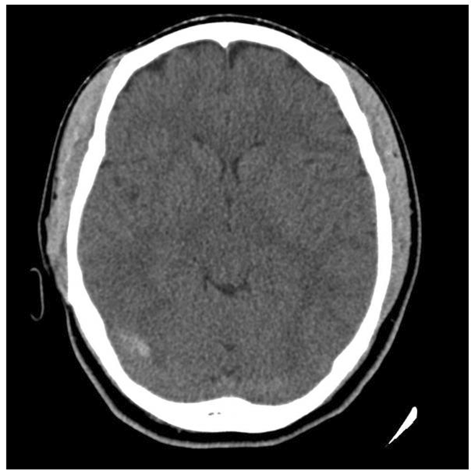
Cerebral Venous Sinus Thrombosis Mrv - VirtualMedStudent.com || Cerebral Sinus Thrombosis / Left lateral sinus thrombosis demonstrated on magnetic resonance venography (mrv).. Valdueza j.m., schmierer k., mehraein s., einhдupl k.m. Assessment of normal flow velocity in basal cerebral veins. Risk factors, presentation, diagnosis and outcome. For this reason, it seems to be mrv allows direct visualization of the dural venous sinuses and the large cerebral veins.19. Cerebral venous sinus thrombosis in children:
Cerebral venous sinus thrombosis created by paul young 22/11/07. 2d time of flight (tof) venography is routinely performed in suspected cases. Secondary sources of data included reference lists of articles reviewed and cohort studies that related treatment to outcomes. Contrast mr venography is more. Cerebral venous sinus thrombosis (cvst) is the presence of a blood clot in the dural venous sinuses, which drain blood from the brain.
Diagnosing pediatric cerebral venous sinus thrombosis ... from www.kjophthal.com Cerebral venous sinus thrombosis (cvst) is less common than other types of stroke but can be more challenging to diagnose due to its varied presentation patterns; H u h n a. Thrombosis involving the superior sagittal sinus, the right transverse sinus, the right sigmoid sinus, and the right jugular vein was diagnosed after neurological deterioration. Efns guideline on the treatment of cerebral venous and sinus thrombosis. Cerebral venous thrombosis (cvt) is a rare form of cerebrovascular disease in which thrombus forms in the dural sinuses and cortical cerebral veins. A summary algorithm for the diagnosis and management of patients with cvt. A statement for healthcare professionals from the american heart association/american stroke association. Assessment of normal flow velocity in basal cerebral veins.
Hanprasertpong t., hanprasertpong j., riabroi k.
Neuroimaging is required for diagnosis, with magnetic resonance venography (mrv) and computed tomography venography (ctv) both being. Thrombosis or cerebral venous thrombosis or sinus thrombosis) and treatment guideline. A summary algorithm for the diagnosis and management of patients with cvt. Cerebral venous thrombosis in the absence of headache. Valdueza j.m., schmierer k., mehraein s., einhдupl k.m. Die thrombosen der intrakraniellen venen und sinus, stuttgart, 1965; Hanprasertpong t., hanprasertpong j., riabroi k. Efns guideline on the treatment of cerebral venous and sinus thrombosis. Assessment of normal flow velocity in basal cerebral veins. 2d time of flight (tof) venography is routinely performed in suspected cases. For this reason, it seems to be mrv allows direct visualization of the dural venous sinuses and the large cerebral veins.19. Cerebral venous sinus thrombosis in early pregnancy: A statement for healthcare professionals from the american heart association/american stroke association.
Cerebral venous thrombosis (cvt) or cerebral venous sinus thrombosis (cvst): This prevents blood from draining out of the brain. Cerebral venous sinus thrombosis (cvst) is less common than other types of stroke but can be more challenging to diagnose due to its varied presentation patterns; The most frequent and often early symptom of thrombosis of cerebral veins and sinuses is a headache. A transcranial doppler ultrasound study.
Cerebral Venous Sinus Thrombosis After Cervical Spine ... from www.journalmc.org Common subtypes include transverse sinus thrombosis and superior sagittal sinus. Neuroimaging is required for diagnosis, with magnetic resonance venography (mrv) and computed tomography venography (ctv) both being. The patient reported that her headache severity was 2/10, with improvement from her chronic baseline state. As a result, blood cells may break and leak blood into the brain tissues, forming a hemorrhage. Cerebral venous thrombosis guidelines 5. Acute symptomatic seizures in cerebral venous thrombosis. Cerebral venous thrombosis (cvt) or cerebral venous sinus thrombosis (cvst): Cerebral venous sinus thrombosis in children:
Left lateral sinus thrombosis demonstrated on magnetic resonance venography (mrv).
Valdueza j.m., schmierer k., mehraein s., einhдupl k.m. For the cerebral venous sinus thrombosis study group. The patient reported that her headache severity was 2/10, with improvement from her chronic baseline state. Cerebral venous sinus thrombosis (cvst) is the presence of a blood clot in the dural venous sinuses, which drain blood from the brain. The average age of the 4 patients in this case series is. Cerebral venous sinus thrombosis (cvst) is less common than other types of stroke but can be more challenging to diagnose due to its varied presentation patterns; Cerebral venous thrombosis (cvt) or cerebral venous sinus thrombosis (cvst): Die thrombosen der intrakraniellen venen und sinus, stuttgart, 1965; This prevents blood from draining out of the brain. The most frequent and often early symptom of thrombosis of cerebral veins and sinuses is a headache. Cerebral venous thrombosis (cvt) refers to occlusion of venous channels in the cranial cavity, including dural venous thrombosis, cortical vein mrv will demonstrate a lack of flow. Subsequent mrv and mra studies showed improved flow of the cerebral venous sinuses. Characteristic, but rare, is the occurrence of unilateral hemispheric.
Review of the scientific literature. Efns guideline on the treatment of cerebral venous and sinus thrombosis. Cerebral venous thrombosis guidelines 5. The patient reported that her headache severity was 2/10, with improvement from her chronic baseline state. Cerebral venous sinus thrombosis (cvst) is the presence of a blood clot in the dural venous sinuses, which drain blood from the brain.
3D High-Spatial-Resolution Cerebral MR Venography at 3T: A ... from www.ajnr.org Characteristic, but rare, is the occurrence of unilateral hemispheric. Cerebral sinus venous thrombosis (csvt) is a rare phenomenon that can be seen with some frequency in young patients. Subsequent mrv and mra studies showed improved flow of the cerebral venous sinuses. Cerebral venous thrombosis (cvt) refers to occlusion of venous channels in the cranial cavity, including dural venous thrombosis, cortical vein mrv will demonstrate a lack of flow. Cerebral venous thrombosis (cvt) or cerebral venous sinus thrombosis (cvst): Thrombosis or cerebral venous thrombosis or sinus thrombosis) and treatment guideline. A statement for healthcare professionals from the american heart association/american stroke association. Common subtypes include transverse sinus thrombosis and superior sagittal sinus.
Cerebral venous thrombosis (cvt) is a rare form of cerebrovascular disease in which thrombus forms in the dural sinuses and cortical cerebral veins.
Review of the scientific literature. As a result, blood cells may break and leak blood into the brain tissues, forming a hemorrhage. In this article, we report an overview of cvst epidemiology, clinical presentation, diagnostics. Thrombosis involving the superior sagittal sinus, the right transverse sinus, the right sigmoid sinus, and the right jugular vein was diagnosed after neurological deterioration. A thrombotic obstruction of the cerebral veins and/or related anatomical structures (dural sinuses) which drain blood from the brain. For this reason, it seems to be mrv allows direct visualization of the dural venous sinuses and the large cerebral veins.19. Common subtypes include transverse sinus thrombosis and superior sagittal sinus. A summary algorithm for the diagnosis and management of patients with cvt. The patient reported that her headache severity was 2/10, with improvement from her chronic baseline state. A transcranial doppler ultrasound study. Efns guideline on the treatment of cerebral venous and sinus thrombosis. Because of the generally good prognosis and variable clinical signs. Barbara simons, geert lycklama a nijeholt and robin smithuis.
Cerebral venous sinus density on noncontrast ct correlates with hematocrit cerebral venous sinus thrombosis. A retrospective case series from pakistan and comparative literature review.


0 Komentar